
What is canine lymphoma? Can it be treated? What is the life expectancy of a dog diagnosed with lymphoma?
Overview of Canine Lymphoma
By Timothy M. Fan, DVM, PhD, DACVIM, Associate Professor, Department of Veterinary Clinical Medicine, University of Illinois
From: Merck Veterinary Manual
Canine lymphoma is a disease term comprising a heterogeneous group of malignancies with varying biologic aggressiveness derived from the uncontrolled and pathologic clonal expansion of lymphoid cells of either B- or T-cell immunophenotype. Although neoplastic transformation of lymphocytes is not restricted to specific anatomic compartments, canine lymphoma most commonly involves organized primary and secondary lymphoid tissues, including the bone marrow, thymus, lymph nodes, and spleen. In addition to these lymphoid-rich organs, extranodal sites affected by lymphoma include the skin, intestinal tract, liver, eye, CNS, and bone. Lymphoma is reported to be the most common hematopoietic neoplasm in dogs, with an incidence reported to approach 0.1% in susceptible dogs. Despite the prevalence of malignant lymphoma, the underlying causes for its development remain poorly characterized; however, advanced genetic studies have revealed that canine lymphoma can be molecularly distinguished and categorized into discrete groups that correlate with biologic aggressiveness. Hypothesized causes include retroviral infection with Epstein-Barr virus–like viruses, environmental contamination with phenoxyacetic acid herbicides, magnetic field exposure, chromosomal abnormalities, and immune dysfunction. With the completion of the dog genome, it is anticipated that genome-wide association studies will identify specific genetic and chromosomal signatures involved in the pathogenesis of lymphoma.
Clinical Findings:
Canine lymphoma is a heterogeneous cancer, with variable clinical signs, responses to therapy, and survival times. The heterogeneity associated with canine lymphoma is influenced in part by several tumor and host factors, including anatomic involvement, extent of disease, morphologic subtype, host constitution, and immunocompetence. In dogs, the most common clinical variants of lymphoma are high-grade T- or B-cell variants, and four conventionally recognized anatomic forms of lymphoma have been described: multicentric, alimentary, mediastinal, and extranodal (renal, CNS, cutaneous, ocular, bone, etc). Multicentric lymphoma is by far the most common anatomic form, accounting for ~80%–85% of all diagnosed cases. The most common and overt clinical manifestation of multicentric lymphoma is the rapid and nonpainful development of generalized lymphadenopathy. In addition to peripheral lymphadenopathy, most affected dogs will have malignant lymphocytes that are detectable by sensitive diagnostic tests, including flow cytometry or PCR for antigen receptor rearrangement (PARR) that involve internal organs, including the spleen, liver, bone marrow, and other extranodal sites. In dogs with significant tumor burden, systemic constitutional signs, including profound lethargy, weakness, fever, anorexia, and dehydration, may become evident.
Alimentary lymphoma accounts for <10% of all canine lymphomas. Dogs with focal intestinal lesions may exhibit clinical signs consistent with partial or complete luminal obstruction (eg, vomiting, constipation, abdominal pain). With diffuse involvement of the intestinal tract, dogs with alimentary lymphoma may show significant and debilitating GI signs, including anorexia, vomiting, diarrhea, hypoproteinemia, and weight loss secondary to malabsorption and maldigestion.
Exclusive involvement of the cranial mediastinum with lymphoma comprises only a small fraction of diagnosed cases; however, sternal lymph node enlargement is frequently observed in dogs with multicentric disease. Mediastinal lymphoma is typically characterized by enlargement of the cranial mediastinal lymph nodes, thymus, or both. Mediastinal lymphoma arising from the thymus is predominantly comprised of high-grade malignant T lymphocytes, and with advanced disease, clinical signs may include respiratory distress associated with pleural fluid accumulation, direct compression of adjacent lung lobes, or caval syndrome. In addition to respiratory signs, some dogs with mediastinal lymphoma may have primary polyuria with secondary polydipsia resulting from humoral hypercalcemia of malignancy, a paraneoplastic syndrome seen in 10%–40% of dogs with lymphoma. Confirmation of humoral hypercalcemia of malignancy can be documented through the measurment of ionized calcium, parathyroid hormone, and parathyroid hormone–related peptide within circulating blood.
The clinical signs associated with various high-grade extranodal lymphomas (which may involve the skin, lungs, kidneys, eyes, CNS, etc) are often variable and dictated by the organs infiltrated. The most common extranodal form of lymphoma involves the skin, referred to as cutaneous lymphoma. Cutaneous lymphoma (epitheliotropic and nonepitheliotropic) may appear as solitary, raised, ulcerative nodules or generalized, diffuse, scaly lesions. Involvement of peripheral lymph nodes and mucocutaneous junctions is frequent. Clinical signs associated with lymphoma involving other extranodal sites may include respiratory distress (lungs), renal failure (kidneys), blindness (eyes), seizures (CNS), and skeletal pain or pathologic fracture (bone).
Although high-grade lymphoma of either B- or T-cell origin is most commonly diagnosed in dogs, low-grade or indolent lymphoma is a recently described molecular variant of canine lymphoma and comprises up to 30% of all lymphoma diagnoses. Like high-grade lymphoma, indolent lymphoma consists of several histopathologic subtypes, including marginal zone, follicular, mantle cell, and T-zone lymphoma. Collectively, indolent lymphomas are slowly progressive, and dogs often remain asymptomatic for a prolonged time regardless of therapy.
Lesions:
Commonly, peripheral and various internal lymph nodes are 3–10 times normal size (multicentric form) and nonpainful on digital palpation. Affected nodes are freely movable, firm, and gray-tan; they bulge on cut surface and have no cortical-medullary demarcation. Frequently, there is hepatosplenomegaly with either diffuse enlargement or multiple, pale nodules of variable size disseminated in the parenchyma. In the alimentary form, any part of the GI tract or mesenteric lymph nodes may be affected. Involvement of the bone marrow, CNS, kidney, heart, tonsils, pancreas, and eyes can be seen but is less common.
Diagnosis:
The definitive diagnosis of lymphoma is often uncomplicated and can be obtained by either cytologic or histopathologic evaluation of the affected organ system. Fine-needle aspiration of enlarged peripheral lymph nodes or affected visceral organs usually provides specimens of adequate cellular content and detail to make a definitive diagnosis. Cytologically, lymph node or tissue aspirates may identify a monomorphic population of lymphoid cells, either of large (lymphoblastic), intermediate, or small size. Despite the ease of diagnosis, conventional cytology is limited for differentiating or categorizing the heterogeneous spectrum of lymphomas with regard to morphologic subtype (diffuse versus follicular, cleaved versus noncleaved) and histologic grade (high versus low). Specialized cytology utilizing lineage-specific antibodies can differentiate between B- and T-cell lymphomas and can provide some information regarding prognosis based on immunophenotype. However, because of the inherent limitations associated with cytology, histopathologic tissue evaluation remains the gold standard for the diagnosis of lymphoma, providing additional morphologic information required for definitive classification, as well as guiding therapeutic decisions.
In rare situations when cytology or histopathology fails to confirm the diagnosis of lymphoma, more advanced molecular techniques are available for definitive diagnosis, including flow cytometry for specific cell surface markers called cluster of differentiation (CD) antigens and PCR for PARR. The use of PCR allows for the amplification of DNA sequences that confirms or denies the presence of lymphocytes of either clonal, oligoclonal, or polyclonal origin. Because most neoplastic outgrowths result from the clonal expansion of one malignantly transformed cell, PCR techniques can differentiate lymphocyte expansion as a consequence of cancer (lymphoma) versus inflammation (reactive or hyperplastic lymphocytosis). Although PCR techniques are highly sensitive, the methodology should be reserved for cases in which conventional cytology and histopathology prove nondiagnostic, or when results are discordant with clinical signs and disease progression.
Treatment:
Treatment of high-grade, multicentric canine lymphoma with aggressive, multi-agent chemotherapy protocols is often rewarding, with >90% of all dogs achieving complete reduction of tumor burden, termed complete remission. The most common chemotherapeutic agents used in combination protocols are vincristine, doxorubicin, cyclophosphamide, l-asparaginase, and prednisone. Individual treatment protocols vary with respect to dosage, frequency, and duration of treatment; advantages and disadvantages of each treatment protocol can be found in medical oncology textbooks. With combination chemotherapy, the expected survival time for dogs with B-cell lymphoma is ~12 mo, whereas for dogs with T-cell lymphoma, expected survival times are often in the range of 6 mo. Although immunophenotype (B- versus T-cell) provides a general guideline for treatment prognosis, multiple factors (tumor and host) contribute to the overall response duration and survival time of dogs diagnosed with lymphoma. Dogs that do not respond to traditional combination chemotherapy or that relapse may achieve disease remission, added survival times, or both with the use of various rescue protocols (eg, lomustine, MOPP, ADIC, DMAC).
Although systemic chemotherapy remains the cornerstone to treat high-grade lymphoma, the idea that both induction and maintenance phases of chemotherapy are necessary to achieve durable remission times has changed. Shorter and more dose-intense chemotherapy protocols (eg, Madison Wisconsin protocol) without maintenance provide disease-free intervals and survival times equivalent to protocols that include chronic maintenance therapy. Additionally, the use of half-body radiation in replacement of maintenance chemotherapy has been demonstrated to be safe and clinically efficacious, providing another option to achieve durable remission times without the need for chronic chemotherapy. For select cases, autologous canine bone marrow transplant after systemic chemotherapy and whole-body radiation can afford some dogs extended, progression-free intervals and survival times.
Despite the favorable outcomes expected in treating high-grade multicentric lymphoma, the successful management of other anatomic forms of lymphoma is often more difficult and less rewarding. Alimentary lymphoma, if focal, can be treated effectively with surgical resection and combination chemotherapy. However, with diffuse involvement of the intestinal tract, low constitutional reserve, severe malabsorption of nutrients, and loss of proteins often result in poor clinical responses and short survival times (ie, <3 mo). The use of combination chemotherapy with or without palliative radiation therapy can afford dogs with mediastinal lymphoma considerable improvement in survival times and quality-of-life scores. Lymphoma involving other extranodal sites (such as the skin) can be managed with either single-agent oral lomustine or combination systemic chemotherapies (eg, CHOP); however, the development of refractory and progressive disease is common and ultimately life limiting.
Clinical prognosis for dogs diagnosed with low-grade, indolent lymphoma tends to be good. The institution of low-intensity oral chemotherapy protocols (chlorambucil and prednisone) often provides prolonged survival times (>2 yr), and in specific dogs with localized and low-grade disease (eg, splenic involvement), splenectomy can be an effective and durable treatment option without the necessity of adjuvant chemotherapy administration. (See also Treatment of Canine Lymphoma.)
Other Sources:
Purdue College of Veterinary Medicine
Canine Lymphomas
Canine lymphomas are a diverse group of cancers, and are among the most common cancers diagnosed in dogs. They collectively represent approximately 7-14% of all cancers diagnosed in dogs. There are over 30 described types of canine lymphoma, and these cancers vary tremendously in their behavior. Some progress rapidly and are acutely life-threatening without treatment, while others progress very slowly and are managed as chronic, indolent diseases. Lymphomas may affect any organ in the body, but most commonly originate in lymph nodes, before spreading to other organs such as the spleen, liver, and bone marrow.
Canine lymphomas are similar in many ways to the non-Hodgkin’s lymphomas (NHL) which occur in humans. Canine lymphomas and NHL are nearly indistinguishable when examined microscopically, and both tumor types exhibit similar responses to chemotherapy. In 2010, NHL was diagnosed in approximately 65,000 people in the United States, and claimed approximately 20,000 lives, making it the 7th-most common cancer overall, and the 6th-most common cause of cancer-related death. It is one of the few human cancers for which the frequency of newly diagnosed cases is still on the rise. It is our hope that research in canine lymphomas conducted by the Purdue Comparative Oncology Program will discover new ways of treating NHL in both dogs and humans. Our goal is to improve the outlook for dogs and humans affected with this all-too-common cancer.
Frequently Asked Questions by Pet Owners
What is lymphoma?
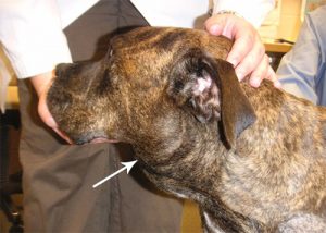
The term “lymphoma” describes a diverse group of cancers in dogs that are derived from white blood cells called lymphocytes. Lymphocytes normally function as part of the immune system to protect the body from infection. Although lymphoma can affect virtually any organ in the body, it most commonly arises in organs that function as part of the immune system such as the lymph nodes, spleen, and bone marrow. By far the most common type of lymphoma in the dog is multicentric lymphoma, in which the cancer first becomes apparent in lymph nodes. The photo to the right shows a dog with multicentric lymphoma. Note the swollen mandibular lymph node (white arrow) under the jaw.
Other common lymphomas in dogs include cutaneous lymphoma (lymphoma of the skin), alimentary or gastrointestinal lymphoma (lymphoma of the stomach and/or intestines) and mediastinal lymphoma (lymphoma involving organs within the chest, such as lymph nodes or the thymus gland).
What causes lymphoma in dogs?
Unfortunately, the cause of lymphoma in dogs is not known. Although several possible causes such as viruses, bacteria, chemical exposure, and physical factors such as strong magnetic fields have been investigated, the cause of this cancer remains obscure. Suppression of the immune system is a known risk factor for the development of lymphoma in humans. Evidence for this includes increased rates of lymphoma in humans infected with the HIV virus or are on immune-suppressing drugs following organ transplantation surgery. However, the link between immune suppression and lymphoma in dogs is not clearly established.
What are the most common symptoms of canine lymphoma?
The most common initial symptom of multicentric lymphoma in dogs is firm, enlarged, non-painful lymph nodes. A lymph node affected by lymphoma will feel like a hard, rubbery lump under your dog’s skin. The most easily located lymph nodes on a dog’s body are the mandibular lymph nodes (under the jaw) and the popliteal lymph nodes (behind the knee). Other common symptoms include loss of appetite, lethargy, weight loss, swelling of the face or legs (edema), and occasionally increased thirst and urination. The photo on the left shows a dog with edema of the left rear leg. This is caused when a swollen lymph node blocks the normal drainage of fluid from the leg.
Cutaneous lymphoma tends to appear first as dry, flaky, red, and itchy patches of skin anywhere on the body. As the disease progresses, the skin becomes moist, ulcerated, very red, and thickened. Masses in the skin can also occur with cutaneous lymphoma. Cutaneous lymphoma may progress slowly and often has been treated for several months as an infection or allergy before a diagnosis of lymphoma is made. Cutaneous lymphoma may also appear in the mouth, often affecting the gums, lips, and the roof of the mouth. Cutaneous lymphoma in the mouth is often mistaken for periodontal disease or gingivitis in its early stages. The photo on the left shows cutaneous lymphoma in the mouth of a dog. Note the very red gums and the ulceration on the roof of the mouth.
Dogs with gastrointestinal lymphoma usually have symptoms such as vomiting, watery diarrhea, and weight loss. The diarrhea is often very dark in color and foulsmelling.
Dogs with mediastinal lymphoma typically have difficulty breathing. This may be due to the presence of a large mass within the chest or due to the accumulation of fluid within the chest (pleural effusion). Affected dogs may also show swelling of the face or front legs as well as increased thirst and urination.
How is canine lymphoma diagnosed?
The best way to diagnose lymphoma is to perform a biopsy. A biopsy is a minor surgical procedure to remove a piece of lymph node or other organ affected by cancer. The most common methods for lymph node biopsy are Tru-cut needle biopsy, incisional wedge biopsy, or removal of an entire lymph node (excisional biopsy). The larger the biopsy sample, the better the chance for an accurate diagnosis of lymphoma.
We routine perform biopsy procedures to diagnose canine lymphoma at the Purdue University Veterinary Teaching Hospital (PUVTH). Dogs are placed under heavy sedation or general anesthesia to perform a biopsy. Although discomfort associated with this procedure is typically minimal, we often prescribe oral pain medication afterwards just to be sure your dog is comfortable following the biopsy.
Are any other diagnostic tests required for dogs with lymphoma?
In addition to biopsy, we recommend several staging tests for dogs with lymphoma. The purpose of the staging tests is to determine how far the lymphoma has spread throughout your dog’s body. In general, the more places the lymphoma has spread to, the poorer the dog’s prognosis. However, dogs with very advanced lymphoma can still be treated and experience cancer remission (see more on treatment below). Staging tests also help us assess whether your dog has any other conditions that may affect treatment decisions or overall prognosis. The staging tests we typically recommend include blood tests, a urinalysis, x-rays of the chest and abdomen, an abdominal sonogram, and a bone marrow aspirate. Organs that appear abnormal on sonogram can be sampled with a small needle (fine needle aspirate) to confirm the presence of lymphoma.
How is canine lymphoma treated?
The most effective therapy for most types of canine lymphoma is chemotherapy. In some cases, surgery or radiation therapy may also be recommended. There are numerous chemotherapy treatment protocols for dogs with multicentric lymphoma. As discussed below, most dogs with lymphoma experience remission of their cancer following treatment, and side effects are usually not severe. Currently, the protocols that achieve the highest rates of remission and longest overall survival times involve combinations of drugs given over several weeks to months. The protocol we use as a “gold standard” for the treatment of canine multicentric lymphoma is a 25-week protocol called UW-25. It is based on a protocol called CHOP that is commonly used to treat lymphoma in humans.
The UW-25 protocol may not be appropriate for all dogs with lymphoma. Different types of lymphoma may be treated with different chemotherapy drugs. For instance, the most effective drug for cutaneous lymphoma is thought to be lomustine (CCNU). The veterinary oncologists and oncology residents at the PUVTH will help you decide on a chemotherapy treatment protocol that is appropriate for your dog.
What does remission mean?
“Remission” means a regression of your dog’s cancer. Remission may be partial, meaning the overall cancer burden has been reduced by at least 50%, or it may be complete, meaning the cancer has become undetectable to any readily available screening test. In general, 70-90% of dogs with multicentric lymphoma treated with UW-25 experience complete or partial remission of their lymphoma, with most dogs experiencing complete remission.
How is chemotherapy given at Purdue?
Most chemotherapy drugs are given by intravenous (IV) injection, although a few are given by mouth as a tablet or capsule. Typically, an IV catheter will be placed in one of your dog’s veins to allow us to administer chemotherapy safely. A small patch of hair will be shaved over your dog’s leg where the catheter is placed.
Chemotherapy appointments with the PUVTH oncology service are on weekdays, Monday – Thursday. Patients are usually dropped off at 9:00 AM and are ready to go home by 12:00-1:00 PM.
Will chemotherapy make my dog sick?
Most dogs tolerate chemotherapy well, much better than humans typically do. Although some dogs do get sick from chemotherapy, serious side effects are uncommon. In general, fewer than 5% of dogs treated for lymphoma using chemotherapy will experience side effects that need to be managed in a hospital setting. The most common side effects include loss of appetite, decreased activity level, and mild vomiting or diarrhea that persists for one or two days. If serious or unacceptable side effects do occur, it is important that you talk to one of our oncology doctors or staff about this. We can recommend symptomatic treatment to lessen the side effects of chemotherapy. In addition we may recommend reducing the dose of chemotherapy the next time it is to be given.
Unlike people, dogs usually do not lose their hair when treated with chemotherapy. The exceptions to this rule are poodles, Old English sheepdogs, and some terriers – these breeds may lose their hair while receiving chemotherapy. Hair growth should resume once chemotherapy is discontinued.
Will chemotherapy cure my dog’s lymphoma?
In rare instances, dogs are apparently cured of their lymphoma by chemotherapy. Unfortunately, most dogs with lymphoma will have relapse of their cancer at some point. A second remission can be achieved in a large number of dogs, but it is usually of shorter duration than the first remission. This is because the lymphoma cells become more resistant to the effects of chemotherapy as time goes on. Eventually, most lymphomas develop resistance to all chemotherapy drugs, and dogs with lymphoma die or are euthanized when the cancer can no longer be controlled with chemotherapy.
What is the prognosis for dogs with lymphoma?
Your dog’s prognosis is determined by what type of lymphoma he or she has and what type of chemotherapy is used to treat the lymphoma. The median length of survival of dogs with multicentric lymphoma treated with UW-25 chemotherapy is between 9-13 months. (The term “median” implies that 50% of dogs will survive beyond this time point and 50% of treated dogs will die before this time point.) Various other factors, such the type of lymphoma your dog has or its stage of disease, may affect your dog’s overall prognosis. The oncologists and oncology residents at the PUVTH will discuss your dog’s prognosis in detail with you before any treatment decisions are made.
Are there any studies at Purdue involving canine lymphoma?
Yes! We are currently conducting multiple clinical trials for dogs with lymphoma at Purdue. Varying degrees of financial support are available to owners who agree to allow their dogs participate in these clinical trials. To determine whether your dog may qualify for a clinical trial, please ask your dog’s primary care veterinarian to call 765-494-1107 and ask to speak with a member of our Canine Lymphoma clinical trials team, or you may contact our Canine Lymphoma Clinical Trials Coordinator, Ms.Sarah Lahrman at 765-496-6289.
Additional Information
Canine Lymphoma: Dogs’ Life Expectancy
Lymphoma in dogs is an aggressive cancer that can metastasize very quickly. If the dog receives treatment and responds well to it, he may live up to one year after the lymphoma is detected. The cancer involves the lymphatic system and spreads at an alarming rate. Surgery is usually not an option, so the prognosis is poor.
Lymphoma in Dogs
Lymphoma may occur as a malignant growth, located in different parts of the body. The cancer will involve the lymphatic system (the lymph nodes). The lymphatic cells may be present in different areas of the body including the skin, stomach, liver or spleen. Lymphoma can start on the skin, bone marrow or an internal organ. The cells will multiply rapidly, affecting neighboring cells and organs, if the condition is not detected and controlled. An early detection of lymphoma and a suitable treatment are decisive in establishing the dog’s life expectancy.
Dog Lymphoma Treatment
Ideally, the lymphoma could be removed through a surgical procedure. However, the cancerous lymphatic cells may be affecting other areas of the body and the surgeon cannot possibly remove all affected lymphatic cells. Typically, surgery is only possible if the cancer is detected very early and is located in a specific area. This is not common, as the dog may not display any symptoms during the early stages of the disease.
The second choice in lymphoma treatment is chemotherapy, which will not eliminate or reduce the number of cancerous cells, but will stop these from spreading and affecting new zones in the dog’s body. Chemotherapy may be combined with different pain medication or anti-inflammatory drugs. Chemotherapy may also be used in conjunction with radiation therapy, which is an efficient pain treatment but will not increase the dog’s chances of survival.
Canine Lymphoma Life Expectancy
The life expectancy for a dog with lymphoma may depend on several factors including:
- The stage of the cancer
- How early the cancer was detected
- Type of treatment administered
- Response to treatment
Canine lymphoma is a forceful cancer and may be fully treated only if surgery is possible. Even if the surgery can be performed, the dog may have other cancerous cells in the body, which will develop further. In the best case scenario, lymphoma can be operated on and the dog can live a normal life after that, while the cancerous cells will never return.
If surgery is not possible and the dog receives chemo drugs, the best prognosis for the dog is to live up to one year, provided the cancer is not metastasized. If the dog doesn’t receive chemotherapy, but rather steroid therapy with prednisone, the dog may live up to six months, but typically will die within two months. If the cancer is already metastasized, the dog has low chances of survival and may live up to four weeks.
Canine Lymphoma Clinical Trials:
https://caninelymphoma.com/canine-lymphoma-clinical-trials/
http://caninecancerawareness.org/clinical-trials-for-canine-lymphoma/1775
http://www.petcancercenter.org/Clinical_Trials_Current_Dogs_Lymphoma.html
https://www.caninecancer.com/clinical-trials/
https://vet-dc.com/resources/animal-cancer-clinical-trials/
http://vetcancersociety.org/pet-owners/clinical-trials/
http://cancer.landofpuregold.com/trials.htm
https://www2.vet.cornell.edu/hospitals/clinical-trials-0
https://www2.vetmed.ucdavis.edu/clinicaltrials/current_trials/by_service/oncology.cfm
For more, Google “Canine Lymphoma clinical trials”