
Bone Cancer — femur
By DR. KARSTEN FOSTVEDT
Bone tumors are malignancies that either begin in bone or spread to bone from other parts of the body. They rarely occur in cats.
Primary bone tumors begin in the bones, and are the most common type. These are called osteosarcomas and chondrosarcomas. Secondary bone tumors, those that spread to bones from other tissues, are much more rare.
Seventy-five percent of primary bone tumors occur in leg bones, but they can also occur in ribs, the spine or the skull. Primary tumors commonly appear in only one location in one bone, whereas secondary tumors often occur in multiple sites in multiple bones.
The cause of bone tumors is unknown; however they can occur at previous fracture sites or sites of previous orthopedic procedures.
Bone tumors usually occur in older, male, large and giant-breed dogs. Dogs with bone tumors usually become suddenly lame. The lameness usually is not associated with trauma. The lameness progresses rapidly to non-weight-bearing, with the dog holding the leg up. There is often swelling of the bone at the tumor site, which can be felt by your veterinarian. There is also muscle atrophy of the affected limb. Common sites of primary bone tumors are the shoulder, wrist and knee.
X-rays will show the classic signs of a bone tumor, which are a combination of bone destruction and bone production. Other diseases, such as fungal infections of the bone, can show similar X-ray signs, so definite diagnosis is always done via bone biopsy. That lets us know what type of tumor is present, and what treatment is needed. Bone tumors commonly spread to the lungs, so chest X-rays are always performed to make sure no spread of the bone tumor has occurred to the lungs.
The main treatment of primary bone tumors is amputation of the affected limb. This is done to relieve the intense pain associated with bone tumors. The surgery rarely cures the bone cancer, because microscopic metastasis has usually occurred by the time the diagnosis has been made. Most dogs do extremely well after amputation.
Bone cancers are extremely malignant, with most dogs surviving only 10-12 months when treated with amputation and chemotherapy.
More on Bone Cancer in Dogs from PetMD
The hallmark of cancer is uncontrolled cell growth, invasion of cells into nearby structures and sometimes a dispersal to distant organs, which is termed metastatic cancer. And since any cell in the dog’s body has the potential to develop into a cancerous cell, bone cancer dramatically illustrates what can happen when things go wrong.
When a cell turns cancerous by a disruption the cell’s physiology, structure or function, normal neighboring cells usually consume the rogue cell. On other occasions the defective cell simply self-destructs and is swept away. But when conditions are just right — or wrong from the animal’s perspective — a modified cell, called a mutant, survives the modification, retains its vigor and reproduces more cells just like itself.
Generation after generation of cells arising from that single mutated cell eventually changes the neighborhood and carves out it’s own territory, spreading its own bad seeds into more and more neighborhoods. Metastatic bone cancer cells break away, hitch hike the blood stream or lymph fluid and travel to entirely new neighborhoods within the dog’s body and begin the malignant process all over again.
Cancer is also termed neoplasia, which means new growth. A cancerous cell grows faster than normal and divides and multiplies at an abnormal rate; its progeny do likewise. From that one abnormal neoplastic cell more like itself invade and crowd out surrounding tissues. With bone cancer, there are four types of cell lines capable of evolving into a neoplastic condition:
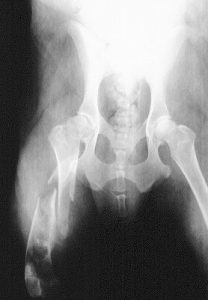
1. Osteosarcoma … causing nearly 80 percent of all bone cancers this most common form of bone cancer arises from cells that deposit bony minerals. Aggressive invasion and rapid growth make this form of cancer a dreaded threat. The X-ray image above shows what an osteosarcoma of the humeral head looks like.
2. Chondrosarcomas … these tumors arise from the cartilage joint surfaces at the ends of bone and generally have a less aggressive tendency to invade and spread.
3. Fibrosarcomas … originate from fibrous connective tissue adjacent to bone, are locally invasive into the bone and have a low tendency to spread.
4. Synovial cell carcinomas … originate from joint tissues and invade the associated bone. These tumors are less aggressive than osteosarcomas.
A definitive diagnosis of bone cancer can only be made via microscopic evaluation of a bone biopsy. Veterinary pathologists classify the degree of malignancy of the cells and likeliness of metastasis to other tissues. Like seeds on the wind, neoplastic cells can be carried by the blood and lymph from the original site of the cancer to distant tissues whereupon a new cancerous growth arises. Called metastatic cancer, whenever distant growths are present in a dog’s body the magnitude of the ill effects on the patient are remarkably increased — and the chances of a cure drastically reduced.
Most commonly seen in long bones such as the femur, bone cancer has a predilection for larger breeds including the Greyhound, Saint Bernard and Mastiff. Chronic, low-grade lameness with gradually increasing swelling near a joint will alert the veterinarian to the potential for a tumor to be present. X-rays of the affected area often display characteristic changes in a bone that are totally unlike the defects usually associated with arthritis.
On occasion, an apparently normal dog will be presented with a spontaneous, severe lameness. Physical examination and radiographic evaluation, to everyone’s shock, reveals the cause of the break to be due to bone cancer. This break is termed a pathological fracture and there is an example of a pathological fracture in the table below.
Osteosarcoma continues to be one of the most challenging types of cancer to treat. Part of the therapeutic challenge arises from the fact that at the time of diagnosis there often has already been metastasis to other areas of the body.
“Unfortunately, at the first signs of lameness,” says Dr. Kenneth M. Rassnick, Assistant Professor of Oncology at Cornell University College of Veterinary Medicine, “we expect that the tumor has already metastasized. However, as long as the metastatic cells are still microscopic and we cannot detect them on radiographs, then dogs will still benefit from treatment.”
There is no single treatment protocol for all patients with bone cancer; Rassnick explains that individualized strategies are selected for each patient. “Currently, for dogs with osteosarcoma, I thoroughly screen them for obvious signs of metastasis. For most dogs, this includes radiographs of the lungs and physical examination and palpation of other bones. Amputation of the affected leg is the first line of treatment but unfortunately, amputation alone is only palliative for a cancer as aggressive as osteosarcoma. Over time, the metastatic cells will continue to grow in number and size. If it is determined by radiography, ultrasound or physical exam that there are no metastatic tumors present, amputation of the affected leg followed by chemotherapy has proved to be the most effective treatment for osteosarcoma. There are a number of chemotherapy regimens that we know to be effective at controlling the metastatic cells.”
Thoughtful consultation with the veterinarian regarding chemotherapy is very important. Rassnick tells us “The exact chemotherapy protocol will depend on a number of factors including overall health status of the dog and function of organs such as the heart and kidneys. We have spent a considerable amount of time trying to determine the best time to begin chemotherapy. Since we know the cancer cells have already spread, it is tempting to begin therapy as soon as possible. Some have even advocated giving chemotherapy before the amputation surgery or even at the same time. Our studies have shown there is not a tremendous benefit to instituting therapy that soon, so I generally recommend having the amputation surgery done, letting my patients heal for 7-14 days and then begin chemotherapy when the stitches are ready to be removed.”
Not all dogs will be candidates for amputation. Rassnick adds “Some dogs may have concurrent orthopedic or neurologic problems that may complicate ambulating with three legs, or occasionally it is the wish of the family not to pursue the surgery. We can offer palliative options for controlling bone pain including nonsteroidal medications and even radiation therapy. Localized radiation therapy to the diseased bone is often a very effective method at controlling the pain and some veterinary oncologists can now offer this as an option.”
As drastic as amputation seems to be, it should not be immediately rejected as an attempt at treatment. As a practitioner for over thirty years I have been amazed at how some canine amputee patients respond and adapt. Each case should be evaluated on its own merits with attention paid to such factors as the presence of arthritis in the patient, degree of excess body weight, heart and other organ function, and the patient’s attitude and ability to adapt to new situations.
Until new research reveals more about the menace of cancer’s mysterious origins and until ways are found to turn off rapidly multiplying cancer cells, we will need to be on the alert for bone cancer in our dogs. A veterinarian should evaluate any lameness that persists longer than three days. All dog owners should be proactive in requesting that an x-ray evaluation be done especially if swelling is present. And no matter what the diagnosis for the lameness is, be certain to let your veterinarian know if the expected healing and return to normal function has not occurred within the expected time frame.
The sooner bone cancer is discovered, the better the chances that treatment will actually affect a cure.
Image: Priority Pet Hospital / via Flickr