
Mast cell tumors are common in dogs. Early detection and staging is critical to diagnoses and successful treatment.
By Catherine Ashe, DVM
From Whole Dog Journal
Mast cell tumors (MCTs) are one of the most frequent skin cancers seen in dogs. Mast cell tumors are the reason why careful monitoring of any skin growths is essential for maintaining a healthy canine. Any new masses on the skin should be evaluated by your veterinarian. In regards to MCTs, there are several predisposed breeds including Boxers, American Staffordshire terriers, and pit bulls.
Mast cells are important in the immune system, particularly in allergic reactions. They are found predominantly in the skin, but they are also found in lower numbers throughout the internal organs. Rarely are they found in the bloodstream. These cells are filled with substances such as histamine and heparin. During an allergic reaction, they degranulate – meaning they empty their contents onto or in the area of the offending allergen. The effect of mast cells can be seen when a patient develops hives and welts, as well as itching and redness.
As with any cell in the body, mast cells can develop cancer. The word cancer merely means the uncontrolled proliferation of cells. It can be divided into 2 broad categories – malignant and benign. Malignant cancers can be locally invasive and damaging, spread to other organ systems like the lungs, or both. Benign tumors do not spread to other organs and are cured by removal.
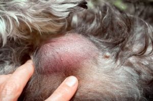
The symptoms of a MCT on dogs begin with a skin mass, most of the time (in rare circumstances, they can start in the internal organs, but this is more common in cats). They can be as small as a pea or as large as a softball. One important aspect is that they tend to wax and wane. They can start as small and suddenly become large, red, and irritated or weepy. This is a sign of degranulation, meaning the tumor has become irritated and released the nasty substances within it. The hallmark of a mast cell tumor is a tumor that grows and shrinks periodically.
If you note a skin mass on your dog, it should be checked by your veterinarian. As with any veterinary visit, your dog should have a nose-to-tail examination including weight and vitals, followed by a detailed history. Your veterinarian will ask how the mass has behaved, how long it has been present, and if it has changed significantly. They may also measure it with calipers.
After a history and physical exam, your veterinarian will focus on the mass with gentle palpation. It is likely they will recommend a fine needle aspirate. This involves taking a very small needle and obtaining a sample of cells from the tumor. Another option is just having the entire mass removed and submitted for testing (excisional biopsy).
If your veterinarian is suspicious of a mast cell tumor and wants to sample it, they may recommend pre-medicating with Benadryl, an antihistamine. As we discussed above, one of the substances found in mast cells is histamine. Giving Benadryl may help prevent the tumor from degranulating during sampling. Sudden degranulation can cause a systemic reaction (anaphylaxis) and can be very serious or even life-threatening. Your veterinarian will handle any suspected MCTs gently, as a result.
Diagnosis is generally by a veterinary pathologist. MCTs are graded on two different scales – the older Patnaik scale (giving a number I through III with I being the least malignant), and the newer Kiupel system that simply evaluates high grade versus low grade.
If your dog is diagnosed with a mast cell tumor, your veterinarian will recommend staging as the next step. This means determining if the cancer has spread by conducting bloodwork, urinalysis, chest xrays, and abdominal ultrasound. Once staging is completed, a clearer picture of prognosis can be seen.
Treatment of mast cell tumors in dogs involves initial surgical removal followed by evaluating whether the whole tumor was removed. If it wasn’t (called “dirty margins”), a second surgery may be needed. Radiation is also an option when the entire tumor wasn’t removed. It may seem “simple” to remove a whole tumor, but sometimes the cancer cells have infiltrated the surrounding tissue on a microscopic level. For low grade (Kiupel system) or grade I-II (Patnaik system), usually removal is sufficient if the margins are clear. Even with removal, a dog will be at higher risk for developing MCTs again.
With high grade/grade III tumors, following surgery, oncologists recommend chemotherapy. This is usually administered by a veterinary oncologist. If chemotherapy is not pursued, a dog with high grade MCT will likely stay on Benadryl and steroids to suppress the MCT until symptoms become too severe. As the disease progresses, the mast cell tumor will spread to distant sites like the liver, spleen, and lungs. Symptoms will correlate with the system that is affected.
A mast cell tumor is not the end of the world, but they can be very serious. It is important to notify your veterinarian when you find any skin masses so that they can be promptly evaluated. Early detection and treatment are critical to a successful outcome.