
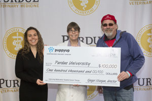
Photo: Dr. Deborah Knapp (center) with Jen Schleicher (left) and the founder of the Puppy Up Foundation, Luke Robinson, at a reception in Lynn Hall celebrating the foundation’s gift of $100,000 for PVM bladder cancer research.
Reception Recognizes Gift to Support Comparative Oncology
January 13, 2017
Purdue Veterinary Medicine’s comparative oncology research program received a major boost in the form of a six figure gift presented just before the winter recess. On December 20, Luke Robinson, founder of the Puppy Up Foundation, stopped by to present a $100,000 gift in support of bladder cancer research being led by Dr. Deborah Knapp, Dolores L. McCall Professor of Comparative Oncology and director of the Purdue Comparative Oncology Program.
At a reception held in the atrium area inside the main east entrance to Lynn Hall, Mr. Robinson shared the story behind the creation of the Puppy Up Foundation, before making the check presentation. He told stories of his adventures with his dogs, including a walking trip from Austin, Texas to Boston, Mass., which inspired him to create the foundation. Now the organization holds fundraising events nationwide during which people bring their dogs to walk two miles in order to raise funds to educate, empower, and invest in comparative oncology research. The collaboration between Dr. Knapp and the Puppy Up Foundation was made possible by a pet owner Dr. Knapp met, Jen Schleicher, who has had a passion for cancer research since losing a dog to bladder cancer in 2015.
During the reception, Dr. Knapp also spoke, expressing her deep appreciation for the Puppy Up Foundation’s support and reviewing progress already achieved through the bladder cancer research being conducted by her team. Click here to view a video in which Dr. Knapp shares more information about PVM’s bladder cancer research program.
Writer: Kevin Doerr, pvmnews@purdue.edu

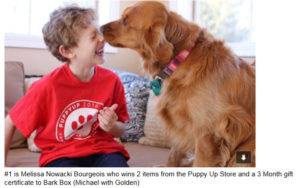
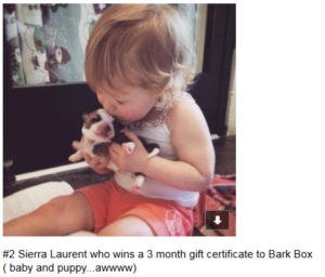
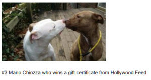
February is about Love. And here at the Puppy Up Foundation to celebrate it, we launched our first ever ‘Puppy Up Kiss Cam Contest.’ People shared photos of their dogs kissing them, kissing other dogs, and even kissing a cow. After much deliberation, a panel of five chose the three contestants below. Congratulations, and thanks for the smooches.
![]()

What IS a Puppy Up Walk? A Puppy Up Walk is a simple 1 to 2 mile easy walk through a park or community for dogs and their owners to enjoy together. Our Walks are a chance for people who might otherwise never meet to get together, share their experiences, and grow a community of people (and dogs) with the same goal – eradicating cancer. The best way we’ve found to do this is through grass roots participation.
Individuals or Teams may walk. Walkers may pay an entry fee to walk, and walkers and teams are encouraged to raise pledges. The top fundraisers will win a prize in each city.
On Sunday, March 26, 2017 from 1 to 5 PM, The Puppy Up Foundation will be having a Walk in Fort Myers, Florida at 6810 Shoppes at Plantation Drive, Fort Myers, FL 33912.
Here’s the schedule:
Registration: 1:30 pm
Opening Ceremony: 2:15 pm
PuppyUp Walk: 2:30 pm
Closing Ceremony: 3:30 pm
And here’s registration information:
Registration: $20.00 Registration after March 24: $25.00
Youth (under 14): Free
Fundraiser: No Fees
For each Puppy Up Walk, the Event Manager and his or her Walk Team select both a Human Cancer Hero and a Canine Cancer Hero, to be honored during the Walk ceremonies.
These Heroes are chosen because of their exemplary attitude towards their particular type of cancer, showing bravery and hopefulness in their fight against this terrible disease.
To learn more about the Walk in Fort Myers, follow this link.
To find out more about ALL the Puppy Up Walks, and how you and your dog can participate, please click here.
Walks are being added around the country. If you don’t see a Walk in your area, please contact us to find out how you can start one in your city or town.

Hemangiosarcoma in Dogs – An Aggressive Blood Vessel Cancer
February 20, 2017 From Critical Care DVM
Many of the people the Puppy Up Foundation has come in contact with throughout the years have told us of the heartbreak of their dogs’ diagnosis of Hemangiosarcoma. This article explains what the disease is, what breeds it most often affects, how it is diagnosed and treated, and new therapies that could benefit your dog. We hope you find this article from Critical Care DVM helpful.
Every pet parent dreads “The Big C” for their fur babies. Cancer strikes fear in the hearts and minds of pet-owning families. As a board-certified critical care specialist, one of the most common cancers I diagnose and treat is called hemangiosarcoma. I wanted to dedicate some time explaining this cancer, as some encouraging news about a novel treatment was recently released.
Hemangiosarcoma – What is it?
Hemangiosarcoma is a cancer of endothelial cells, the cells that line blood vessels. We don’t currently know what causes some dogs to develop this cancer. Multiple studies have tried to shed more light on this topic. Risk factors include:
· Heritability – certain breeds are over-represented for developing this cancer
· Ultraviolet light exposure – long-term exposure in lightly pigmented short-haired breeds increases risk
· Abnormal gene expression
· Abnormal development of new blood vessels
Hemangiosarcoma usually occurs in middle-aged and geriatric dogs. Certain breeds are over-represented, including:
· Golden retrievers
· German shepherds
· Labrador retrievers
· English setters
· Boxers
· Doberman pinschers
Hemangiosarcoma can develop anywhere in the body where there are blood vessels. This cancer is often classified as follows:
· Dermal – The skin form typically appears as red or black skin growths. These masses can become ulcerated and bleed. Approximately 33% of these tumors will spread to internal organs, so early identification and removal are key.
· Subcutaneous – The layer immediately beneath the skin – called the subcutaneous tissue or hypodermis – can develop dark red to black growths even though the overlying skin looks completely normal. Pet parents simply feel a lump beneath the skin. Almost two thirds of subcutaneous hemangiosarcoma spreads internally.
· Visceral – Hemangiosarcoma affecting internal organs or viscera is the most common manifestation of this aggressive cancer. The most commonly affected organs are the spleen, heart, and liver. Visceral hemangiosarcoma if often life-threatening since tumors tend to rupture and bleed profusely.
Hemangiosarcoma – How is it diagnosed?
Pet parents should regularly check their fur babies for skin masses.
Any skin mass larger than a pea (1 cm) and/or any that has been present for more than one month should be evaluated by a veterinarian.
The doctor should aspirate some cells from the mass(es) to determine the type of growth. No one – not even a veterinarian – can simply look at a skin mass and know what it is. As DrSueCancerVet advocates, #WhyWaitAspirate! Early identification and intervention for the dermal and subcutaneous forms of hemangiosarcoma are of paramount importance!
Patients with visceral hemangiosarcoma often don’t have any clinical signs until a tumor ruptures to cause internal bleeding. Depending on the location and severity of the bleeding, patients may be depressed, refuse to walk or play, develop pale (or white) gums, have trouble breathing, collapse, and/or have a distended abdomen.
A veterinarian should obtain a thorough patient history and perform a complete physical examination. Initially, some blood and urine tests, as well as some diagnostic imaging, will be recommended:
· Complete blood count – a non-invasive blood test that measures red blood cells, white blood cells, and platelets
· Serum biochemical profile – a non-invasive blood test that assesses liver and kidney function, as well as electrolytes (i.e.: sodium and potassium) and certain gastrointestinal enzymes.
· Urinalysis – non-invasive urine test that helps to evaluate renal function and to screen for possible urinary tract inflammation and infection.
· Coagulation profile – a non-invasive blood test helps determine if a patient can properly form a proper blood clot
· Chest radiography (x-rays) – a non-invasive imaging test to screening for evidence of heart and lung changes, including metastasis (cancer spread)
· Abdominal radiography +/- sonography – non-invasive imaging of the abdomen can assess organ size and architecture, and identify tumors of internal organs
· Echocardiography – Sonographic examination of the heart can evaluate this organ’s function, identify tumors of the heart, and confirm the presence of abnormal fluid in the sac around the heart (called pericardial effusion) caused by a bleeding tumor
A definitive diagnosis of hemangiosarcoma is made by removal and biopsy of the tumor.
Hemangiosarcoma – How it is treated?
Treatment depends on the location of the tumor. When identified early, surgical removal of dermal hemangiosarcoma can be curative. The reported median survival time for the dermal form is 780 days. Chemotherapy may be recommended if the skin tumor couldn’t be completely removed or if it spread to the underlying subcutaneous tissue. Similarly, subcutaneous hemangiosarcoma should be resected whenever possible. Unfortunately, complete excision is not often possible. Therefore, patients with this form of disease may benefit from chemotherapy and/or radiation therapy. The medial survival time for subcutaneous hemangiosarcoma is 172-307 days.
Visceral hemangiosarcoma requires aggressive therapies. Patients with bleeding masses in internal organs generally need emergency surgery. The spleen is not a vital organ, and can be surgically removed. Similarly, bleeding tumors of the liver can often be removed. Unfortunately, not every tumor of an internal organ can be excised, and some patients have inoperable disease. Bleeding into the sac around the heart (called the pericardium) also requires immediate intervention. The fluid must be removed via a procedure called a pericardiocentesis so the heart can function properly. Family veterinarians often refer families to veterinary specialty hospitals where board-certified veterinary surgeons and emergency/critical care specialists can provide life-saving interventions and post-operative critical care.
Surgery alone unfortunately doesn’t appear to confer a marked survival benefit for patients with visceral hemangiosarcoma. The reported median survival times for dogs with splenic hemangiosarcoma treated only with surgery are 19-86 days. Nevertheless, patients who do undergo surgery tend to feel better in the short term.
Chemotherapy after surgery is often recommended because hemangiosarcoma is highly malignant and readily metastasizes. Indeed, this cancer has typically already spread at the time of diagnosis. Many different chemotherapeutic agents have been investigated to treat hemangiosarcoma. Use of the drug doxorubicin is associated with longer survival times. The reported median survival times for splenic hemangiosarcoma treated with surgery and doxorubicin-based chemotherapy is 141-179 days. The prognosis for heart-based hemangiosarcoma is very poor. Families should consult with a board-certified veterinary oncologist to ensure the most appropriate treatment plan is selected for their fur baby.
Hemangiosarcoma – Are there any new treatments?
Recently some novel interventions have been reported to afford a survival benefit in dogs with specific forms of hemangiosarcoma:
· Antibody therapy – Cancer researchers at the University of Pennsylvania’s School of Veterinary Medicine are investigating the efficacy and safety of treatment with antibodies to inhibit the activity of vascular endothelial growth factor or VEGF. This protein stimulates angiogenesis, the formation of new blood vessels. Dysregulated formation of new blood vessels has been implicated as a cause of hemangiosarcoma.
· Immunotherapy – Liposome-encapsulated muramyl tripeptide phosphatidylethanolamine (L-MTP-PE) has been used in combination with splenectomy and systemic chemotherapy for the treatment of hemangiosarcoma. Results of study published in the mid 1990s showed L-MTP-PE conferred a significant survival benefit in dogs with splenic hemangiosarcoma. Unfortunately, this therapy has not been approved in the United States and is not commercially available now.
· I’m-Yunity – This is a newer compound derived the mushroom, Coriolus versicolor. A bioactive chemical in the mushroom called polysaccharopeptide has antitumor activities and inhibits the growth tumors in animal models. In a recent study performed at the University of Pennsylvania’s School of Veterinary Medicine, I’m-Yunity significantly improved survival times in dogs with splenic hemangiosarcoma compared to those who only underwent surgery to remove the spleen (199 days vs. 86 days). This same oncology team is now currently conducting a clinical trial using I’m-Yunity to assess the efficacy and side effects of I’m-Yunity compared to doxorubicin-based chemotherapy after splenectomy.
· eBAT – Earlier this month a team of researchers and board-certified veterinary oncologists at the University of Minnesota’s College of Veterinary Medicine published exciting results in the journal Molecular Cancer Therapeutics. They reported on the effects of a new drug called eBAT used in dogs with splenic hemangiosarcoma. This new drug improved the 6-month survival rate to approximately 70%. Additionally, 21% lived more than 450 days! This is an exciting breakthrough, and further use is need to corroborate these initial encouraging results.
The take-away message about hemangiosarcoma in dogs…
Hemangiosarcoma is a highly malignant cancer arising from endothelial cells. Tumors may be found anywhere in the body, and most commonly affects the spleen, liver, heart, and skin. Early identification and treatment are essential. Treatment may involve surgery, chemotherapy, and radiation therapy. Recently, some novel interventions have been investigated, and preliminary results are encouraging.
To find a board-certified veterinary oncologist, please visit the American College of Veterinary Internal Medicine.
To find a board-certified veterinary surgeon, please visit the American College of Veterinary Surgeons.
To find a board-certified veterinary emergency and critical care specialist, please visit the American College of Veterinary Emergency and Critical Care.
![]()
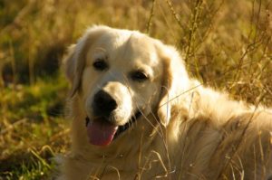
Angel Warner
Angel
(by Michelle Warner) Angel was her name and she lived up to it. This picture is a few weeks before she was taken from us. We had adopted her after our last dog Ruby had died of hemangiosarcoma. We were on vacation in the Smoky Mountains and Angel just wasn’t eating like usual. We thought it was because she was away from home in a new place even though she was with us. We got back home and she still didn’t want to eat. We took her to the vet and at first they couldn’t find anything wrong and gave her some meds thinking she may have picked up something. For a day she seemed a little bit better but then she took a turn for the worse. Took her back to the vet and with ultrasound they found she had liver cancer. We were devastated since we had only had Angel with us for 18 months. She was only 4. There was nothing they could do so we had to let her go.
(Angel was a contestant in the 2014 Puppy Up Calendar Contest.)